
The Magnetic Resonance Imaging (MRI) framework (D5.1) to simulate cardiac metabolism, MRI pulse sequences, image reconstruction and data fitting to provide means for MRI sequence optimization and sensitivity analysis has been successfully accomplished (Figure 1). The work has been presented at the recent Annual Meeting of the International Society of Magnetic Resonance in Medicine in Montréal (www.ismrm.org/19m).
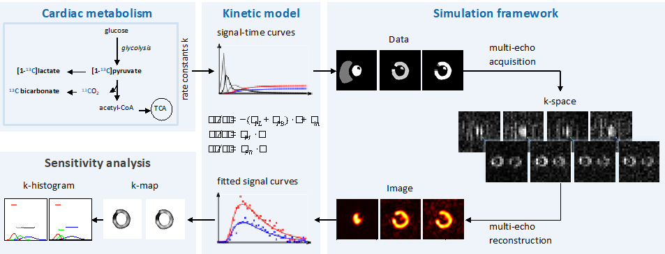
MRI simulation framework including modeling of cardiac metabolism using kinetic models to generate signal-time curves, which feed into data encoding and sampling for subsequent image reconstruction, which in turn allows data fitting to retrieve metabolic parameters. The framework permits sensitivity analysis and hence optimization of MRI metabolic imaging protocols (Traechtler J et al.).
In view of the limited “lifetime” of hyperpolarized states when using e.g. [1-13C] pyruvate, work regarding the hyperpolarization of “long-lived” spin states has been continued. Using suitable 29Si nanomaterials, longitudinal relaxation times (T1) of more than 30 min are achieved (compared to e.g. 30-50 sec for [1-13C] pyruvate).
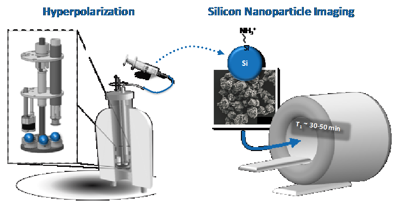
Generation of “long-lived” hyperpolarization using 29Si nanomaterials. Using home-built experimental hyperpolarization equipment (left), angular momentum of silica surface electrons is transferred to 29Si nuclei achieving ~10% polarization with a lifetime of hours.
As part of explorative work, the feasibility of MRI of hyperpolarized substrates at very low magnetic fields has been simulated (Figure 3). This side project is motivated by endeavors to provide ubiquitous MRI diagnostic. Given the cost involved in siting strong magnets as in clinical MRI, hyperpolarized imaging may offer routes to less bulky and hence more cost-effective imaging.
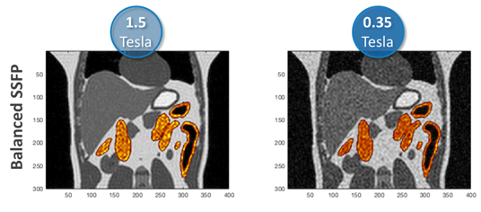
Simulation of MRI of hyperpolarized 29Si nanomaterial at a clinical 1.5 Tesla MRI system (left) and at a hypothetical low-cost 0.35 Tesla MRI system (right). While background image data at low field is more noisy, the hyperpolarized substrate (yellow) is still imaged with sufficient signal-to-noise ratio.